Not Every Sublingual Mass in a Cat Is Cancer: Key Rule-Outs
A one-year-old ragdoll's SCC-mimicking sublingual swelling proved to be an eosinophilic granuloma resolved with two weeks of steroids. Ranulas, foreign bodies and more belong on the rule-out list.
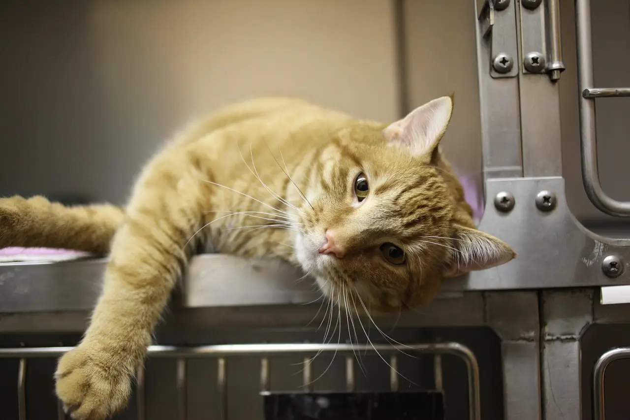
Photo: bongbabyhousevn / pixabay
A one-year-old ragdoll cat named Milo presented with a firm, verrucous, ulcerated swelling beneath the tongue that convincingly mimicked squamous cell carcinoma — and histopathology proved it was an eosinophilic granuloma that resolved fully with two weeks of steroid therapy. The case, reported by John Lewis, VMD, DAVDC, FF-OMFS, of Veterinary Dentistry Specialists in Chadds Ford, Pa., is a reminder that betting on the odds carries a cost: a 10-year-old cat with bloody drool, reduced appetite and dysphagia makes sublingual SCC the statistical favorite, but several less common differentials deserve a place on the list before treatment decisions follow.
What belongs on the differential list?
- Ranulas — smooth, fluctuant sublingual swellings from saliva accumulating after damage to the mandibular or sublingual ducts or their caruncle openings.
- Eosinophilic granulomas — firm, sometimes ulcerated swellings that can mimic either SCC or a ranula.
- Linear foreign bodies — string or twine wrapped around the lingual frenulum, causing edema, erythema and tongue swelling.
- Granulation tissue — a chronic irritation response, for example from the rough surface of a resorbing tooth.
- Pseudoranulas — postoperative sublingual swelling and edema after duct transection during mandibulectomy, which resolve within days.
Each of these changes what happens at the exam table: biopsy, not assumption, drives the diagnosis.
How do ranulas form?
The name comes from the Latin rana, frog, because the swelling resembles a frog's underbelly. Lift the tongue of a cat or dog and you will see paired raised caruncles near the midline of the caudal symphysis. These caruncles are the duct openings for the mandibular and sublingual salivary ducts. Damage to the caruncles, or to the ducts running between glands and caruncles, lets saliva extrude into the tissue.
The resulting swelling may be unilateral or bilateral and is typically not inflamed, though a large swelling can suffer secondary trauma from the teeth. Ranulas occasionally resolve without treatment. More often, surgery is required to remove the mandibular and sublingual glands.
An experimental study from 1978 (DeYoung, Kealy and Kluge, Am J Vet Res, 39:185-186) showed that surgical trauma to the duct or gland did not always produce salivary accumulation — no ranulas developed in those dogs, though the sample was small. Lewis notes he has seen at least one ranula arise postoperatively after duct transection during oncologic surgery, and postoperative pseudoranulas after mandibulectomy typically resolve within a few days.
The takeaway: duct injury alone does not guarantee a ranula, but it remains a real postoperative possibility.
Why did Milo's case matter?
Eosinophilic granuloma is classically associated with rodent ulcers on the upper lip, but not all manifestations are ulcerated. Lip or sublingual swelling belongs on the eosinophilic differential list, and a sublingual eosinophilic granuloma can look strikingly smooth — exactly like a ranula.
Kirk Herrmann, DVM, DAVDC, Alex Juriga, DVM, and Lewis managed Milo jointly. The swelling was so severe that, while awaiting biopsy results, they worried about self-trauma from the teeth. They started dexamethasone sodium phosphate, then oral prednisolone at 1 mg/kg/day beginning the following day. The swelling decreased significantly within one week and was nearly resolved after two weeks of steroid therapy.
Histopathology of the sublingual lesion showed marked eosinophilic granulomatous glossitis with ulceration. A swollen lower lip, still slightly enlarged at presentation, supported the eosinophilic diagnosis; a previous cat in Lewis's series had a firm, non-fluctuant sublingual swelling plus lip swelling, both confirmed by incisional biopsy and resolved with steroids. The team recommended a dermatology consult to pursue underlying allergic causes.
The takeaway: histopathology converted a presumed cancer into a steroid-responsive disease.
Which other rarities should clinicians catch?
Linear foreign bodies wrap around the frenulum and cause significant sublingual edema, erythema and tongue swelling. They occur most often in young cats but also in dogs: Lewis recalls Lily, a 10-year-old dog that chewed the inner twine of a golf ball, which wrapped around the base of the tongue. Chronic irritation, such as the rough surface created by tooth resorption, can also produce a large area of sublingual granulation tissue.
The takeaway: age and signalment shift the odds, but they never close the list.
For practitioners, the practical next step is straightforward: when a sublingual lesion appears in a cat, run the full differential and biopsy before committing to a prognosis — and expect future cases like Milo to respond quickly when the diagnosis is right.
Source: Veterinary Practice News
Elena Vasquez
Show full bio
Market editor covering money, funding and market shifts around Small- and large-animal clinical practice at The Vet Scope.
Related articles
Feline Chronic Rhinosinusitis: New Review Maps Diagnostics and Limits of Antibiotics
WSAVA Releases 2026 Global Dental Guidelines for Pets
Feline BAL Case Puzzle: Wiley Publishes New Diagnostic Exercise
Cornell Identifies Rare Human Genetic Disorder in Two Cats
DVM360 frames feline panleukopenia from the diagnostic bench