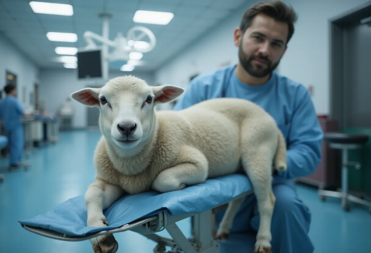
Obstructive Urolithiasis in Small Ruminants: Diagnostics and Treatment
Ultrasound bladder cutoffs of 8 cm and 6 cm, urethral process amputation in every case, and 20–55% postoperative complication rates define current obstructive urolithiasis management in small ruminants.
Updated
A bladder diameter greater than 8 cm on ultrasound indicates obstruction in large-breed goats such as Boer or Nubian; in smaller breeds like Nigerian Dwarf or Pygmy, the cutoff is 6 cm. Those numbers come from a clinical review of obstructive urolithiasis in small ruminants, an emergency the authors say demands swift diagnosis because complete obstruction can cause life-threatening electrolyte imbalances, permanent renal damage, and bladder or urethral rupture.
The condition is increasingly common as goats and sheep gain ground as companion animals. Castrated males are over-represented, likely because early castration leaves a narrower urethral lumen. Common obstruction sites are the two curves of the sigmoid flexure and the urethral process at the distal tip of the penis.
Which signals should trigger urgent workup?
Owners may report straining to urinate, urine dribbling, straining to defecate, or apparent constipation. Exam findings include:
- Tail flagging, vocalizing, flank watching, and a stretched-out stance
- Tense or distended abdomen
- Lethargy or inability to stand in severe cases
- Bradycardia or bradyarrhythmia, which signals hyperkalemia and requires immediate intervention
- Crystalline debris on the hair around the prepuce
- Ventral abdominal swelling or edema suggesting urethral rupture
Timeline drives prognosis. An animal showing clinical signs for less than 24 hours is more likely to have a positive outcome, making an accurate history a determining factor.
What do ultrasound and radiographs change at the table?
Ultrasonography is the most useful tool for confirming obstruction — a round, turgid bladder is diagnostic regardless of diameter. Unlike in small animals, uroliths themselves are often not visible on ultrasound in ruminants, though a hyperechoic "snow globe" appearance of urine suggests struvite crystalluria. Free abdominal fluid indicates uroperitoneum; if no defined bladder is visualized, suspect rupture, which calls for emergency surgery or euthanasia.
Radiography suggests urolith type and location. Rounded mineral-opacity stones point to calcium-based uroliths — a critical distinction, because calcium carbonate uroliths require surgical removal. Struvite and other phosphatic stones are rarely visible in larger patients but may appear in smaller ones. Contrast studies help when stones are not detected or urethral stricture or rupture is suspected.
On bloodwork, abnormal packed cell volume, severely increased creatinine, and elevated creatine kinase have been associated with increased risk of nonsurvival. Hyperkalemia from prolonged obstruction or bladder rupture can produce life-threatening bradyarrhythmia.
What does medical management involve?
Urethral process amputation is both diagnostic and therapeutic and should be performed in all cases. After sedation and exteriorization, an 8- or 10-Fr polypropylene catheter can be passed retrograde to assess patency and flush debris; full passage to the bladder is usually impossible because the catheter diverts into the urethral recess.
Percutaneous transabdominal catheters — Bonanno, pigtail, or trocar-placed Foley — provide temporary diversion. One study described patency of only 1 to 4 days, but that window can allow struvite dissolution or resolution of urethral inflammation. Blockage and migration are common complications.
Only struvite uroliths dissolve with urine acidification. The goal is a urine pH below 6.5 for 4 to 5 days, using ammonium chloride or ᴅʟ-methionine orally, or direct Walpole's solution infusion through the catheter. Pulse dosing — 3 days on, 4 days off, repeated 2 to 3 times — helps patients needing prolonged therapy. Long-term routine acidification is discouraged: animals develop tolerance, and prolonged ammonium chloride use increases urinary calcium excretion, potentially promoting calcium-containing stones.
What are the surgical options?
Tube cystotomy under general anesthesia places a Foley catheter (8 to 28 Fr by patient size) secured in the bladder for 10 to 14 days, allowing urolith removal, lavage, and urethral flushing. Broad-spectrum antibiotics may be warranted given ascending infection risk. If the patient fails a repeated Foley occlusion challenge, salvage procedures or euthanasia should be discussed.
Perineal urethrostomy suits patients with repeated obstructions or distal penile rupture, but stricture is common and obstruction often recurs within 1 to 2 months. The modified technique may reduce stricture risk but carries higher hemorrhage rates. The procedure eliminates reproductive ability. Bladder marsupialization and vesicopreputial anastomosis remain last-resort options requiring significant follow-up for cystitis and urine scald.
What should clients hear about recurrence?
Studies show 20% to 55% of postoperative patients experience complications, with reobstruction the most common; a 2021 study found a mean time to first postoperative complication of approximately 6 months. Prevention centers on adequate water intake and a dietary calcium-to-phosphorus ratio of 2–3:1. Delaying castration until 6 months of age may maximize urethral growth, though intact males still obstruct and some animals form stones with no identifiable risk factors.
Practices treating small ruminants should also note that extrabel drug use — sedatives, muscle relaxants, NSAIDs such as flunixin meglumine and meloxicam, and antibiotics — requires FARAD consultation for meat and milk withdrawal times. As the review emphasizes, removing the obstruction is treatment, not cure; recurrence and prognosis discussions, including euthanasia as a humane endpoint, belong in every discharge conversation.
Original: go.navc.com
Grace Kim
Show full bio
Staff writer covering regulation, policy and their impact on Small- and large-animal clinical practice at The Vet Scope.
Related articles
Neurologic disease in goats: a systematic pathway to diagnosis
Thoracic Ultrasound Evaluated as Diagnostic Tool for Sheep Respiratory Disease
Nanofibrillated cellulose tested as urethral bulking agent in dogs
Thoracic Ultrasound Assessed as CCPP Diagnostic in Sheep and Goats
High-Protein Grazing Raises Blood Ammonia but Not Summer Scour Risk