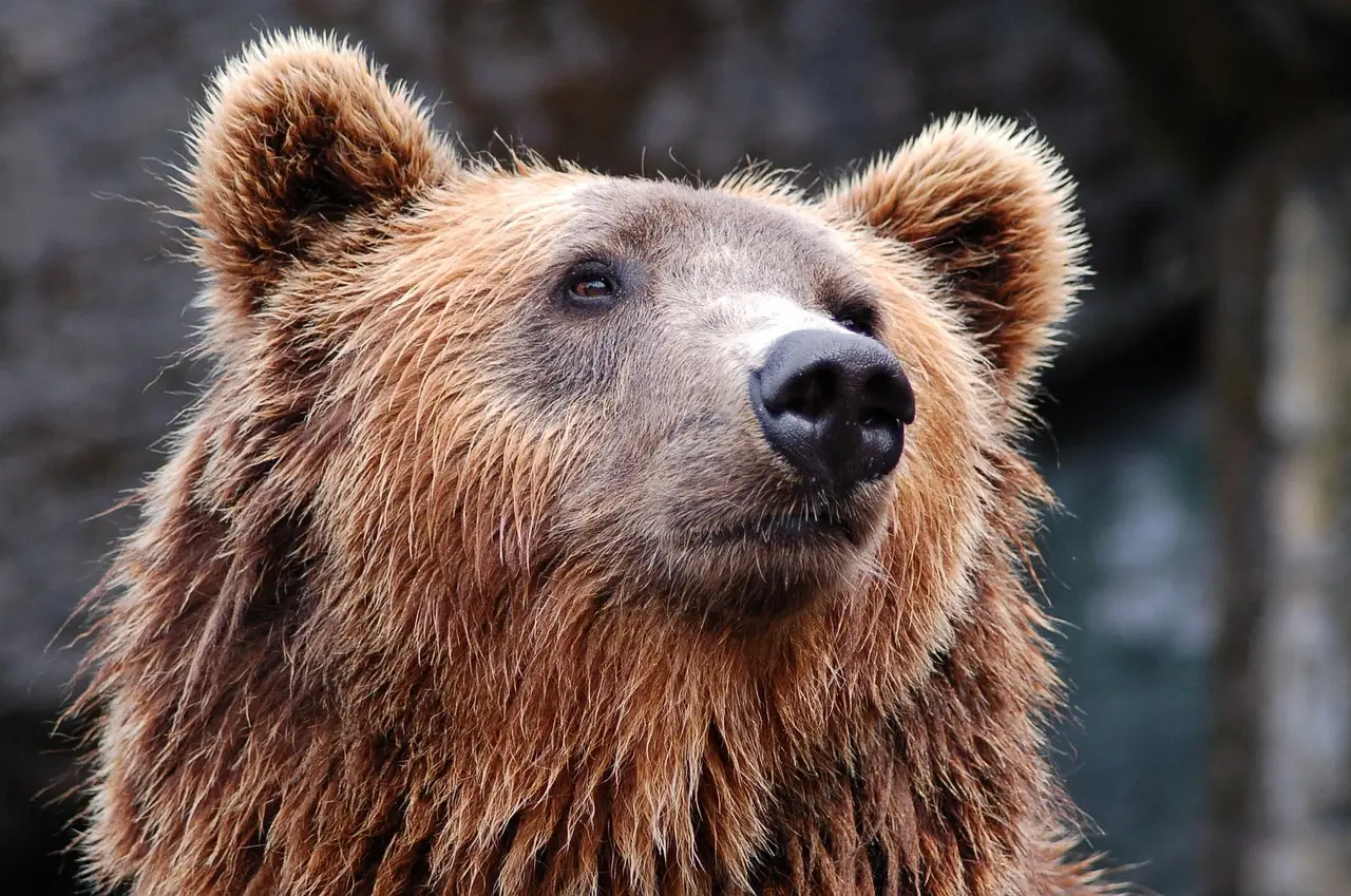
500-pound grizzly Adak undergoes two-hour root canal at WSU
An 11-year-old, 500-pound grizzly received a two-hour root canal at WSU after a canine tooth fracture exposed pulp, preserving function without extraction.
Photo: Pexels / pixabay
By Amara Osei
A 500-pound, 11-year-old grizzly named Adak underwent a roughly two-hour root canal at Washington State University’s Veterinary Teaching Hospital after a fractured canine exposed the tooth’s pulp.
The report describes one animal at WSU’s Bear Research, Education, and Conservation Center in Pullman, Washington, and functions as a single-animal clinical account rather than a comparative study.
Chelsea Davis, the Bear Center’s animal care facilities manager, said: “We suspect Adak broke off the tip of his tooth while wrestling with his sister.” Adak had already been scheduled for an anesthetic procedure the next day, so Davis coordinated with Gay Lynn Clyde of Campus Veterinary Services for a more detailed examination while he was anesthetized.
The exam confirmed exposed pulp. Davis then coordinated with the Veterinary Teaching Hospital and Raelynn Farnsworth, College of Veterinary Medicine associate dean and professor, to schedule the root canal. Farnsworth teaches animal dentistry at WSU.
Why did the Bear Center refer Adak?
Davis said a root canal was beyond the Bear Center’s capabilities and that its partnership with the veterinary hospital improves the likelihood of successful dental procedures. She described tooth problems as the number one health problem for bears.
Sparring and chewing enrichment items such as bones and logs can wear bear teeth, Davis said. Early detection gives the center a good chance to preserve the rest of a damaged tooth and support better future health.
How did the team perform the root canal?
Beforehand, Davis and colleagues gave Adak a pre-treatment medication and kept him fasting. Veterinarians then anesthetized the 500-pound animal and moved it across campus in an animal transport van to the Veterinary Teaching Hospital.
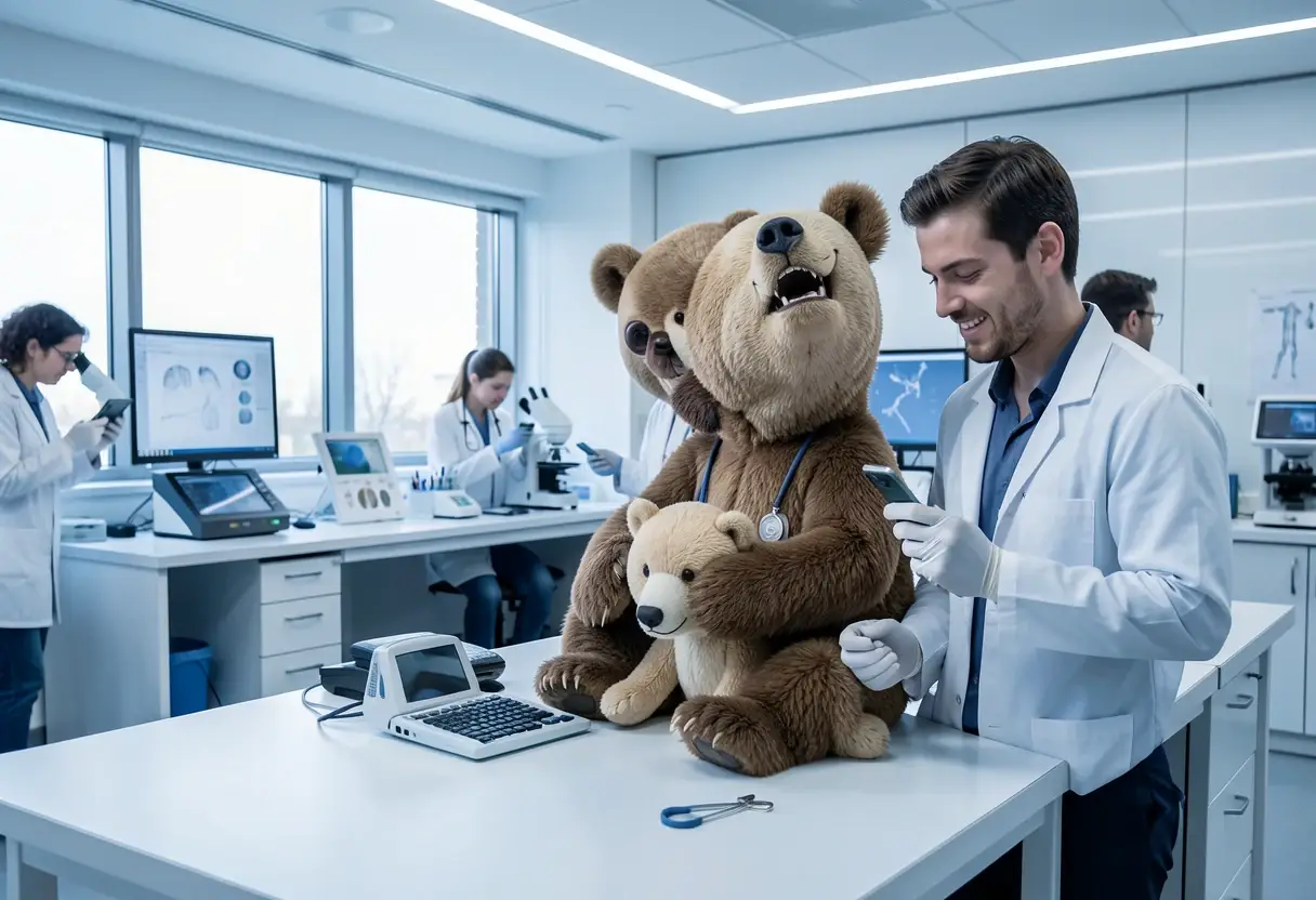
The clinical group included a boarded veterinary dentist, a boarded anesthesiologist, a fourth-year WSU veterinary student, Farnsworth and several graduate students. Davis said the Bear Center trusts the hospital team with its animals.
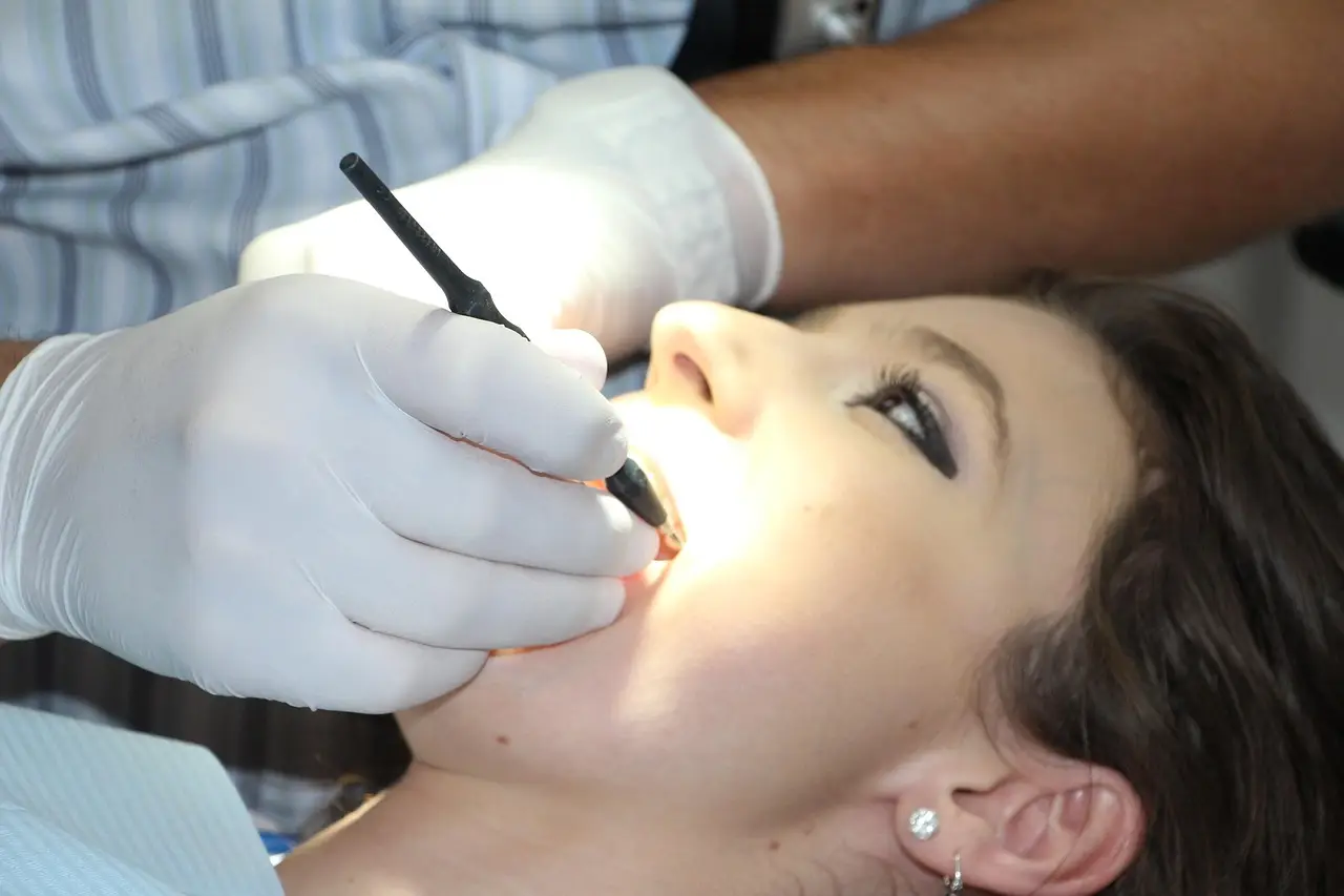
The team first radiographed the tooth’s root, numbed the tooth and drilled an access hole. They cleaned out the live portion containing the tooth’s nerves and blood supply, sterilized the root and inserted an inert substance into the canal to prevent infection.
A second radiograph confirmed that the root was completely filled. The veterinarians then placed a filling over the access hole. The entire procedure took roughly two hours.
What happened after the procedure?
The team gave Adak a long-lasting numbing medication for recovery, transported him back to the Bear Center and allowed him to wake in his den. As part of routine care, he also received pain medication and antibiotics.
Adak’s tooth was not infected. “Luckily Adak’s tooth wasn’t infected, but we still took care of it as quickly as possible because we can’t just put the bears under anesthesia whenever we want,” said Farnsworth. “It must be timed appropriately so they’re comfortable.”
Farnsworth said long-term oral health affects eating, appetite and infection risk in bears. Prompt treatment also matters because the center cannot anesthetize a bear whenever it chooses.
How did the root canal differ from extraction?
According to Davis, extraction usually requires medication, continual follow-up and eight to 12 weeks of diet modification. Adak could return immediately to his normal diet after the root canal.
Davis said the primary benefit is preservation of the tooth and its function. A functional tooth supports normal chewing and feeding and, over time, benefits the animal’s overall health; the center prefers saving a tooth to removing it when possible.
What does this case mean for other animals?
For the Bear Center, the direct lesson is to use scheduled anesthesia to examine suspicious teeth early and refer suitable cases for specialist dental treatment. The report does not establish outcomes for other bears or wildlife populations.
- Companion animals: The report provides no new evidence for dogs or cats and no prescribing information that would change companion-animal dentistry.
- Food animals: No food-animal findings appear in the case, so it offers no basis for changing herd dental or medication protocols.
- Public health: The report identifies no zoonotic-disease finding, outbreak or public-health recommendation and makes no food-safety claim.
The case identifies four medication categories but no products: a pre-treatment medication, a long-lasting numbing medication, pain medication and antibiotics. It supplies no active ingredients, doses or routes, reports no FDA CVM, EMA or USDA regulatory status for any product, and mentions no vaccine.
The collaboration also served as a teaching case. Raj Khosla, Cashup Davis Family Endowed Dean of the College of Agricultural, Human, and Natural Resource Sciences, characterized the work as a prime example of cross-college partnership and praised the staff and faculty for keeping Adak comfortable.
Farnsworth said the team rarely performs root canals, making the procedure a chance to preserve a functional tooth that supports normal bear behavior and show veterinary students that expertise has limits and collaboration is central to animal care. A video of the procedure is also available from WSU.
For future cases, Davis and Farnsworth’s approach points to one defined pathway: inspect a damaged tooth during planned anesthesia, refer root-canal candidates to the teaching hospital and preserve function before infection develops.
Source: Google News: Veterinary medicine
Amara Osei
Show full bio
Correspondent covering companies, deals and strategy in Small- and large-animal clinical practice at The Vet Scope.
Related articles
WSAVA Releases 2026 Global Dental Guidelines for Pets
Millions of Fibers, One Extraction: The Instruments That Do the Work
University of Tennessee vets treat black bears at 'fix-a-bear workshop'
Surgeons Reconstruct Border Collie's Skull After Horse Kick
UC Davis Reappoints Stetter as Dean of Veterinary Medicine School