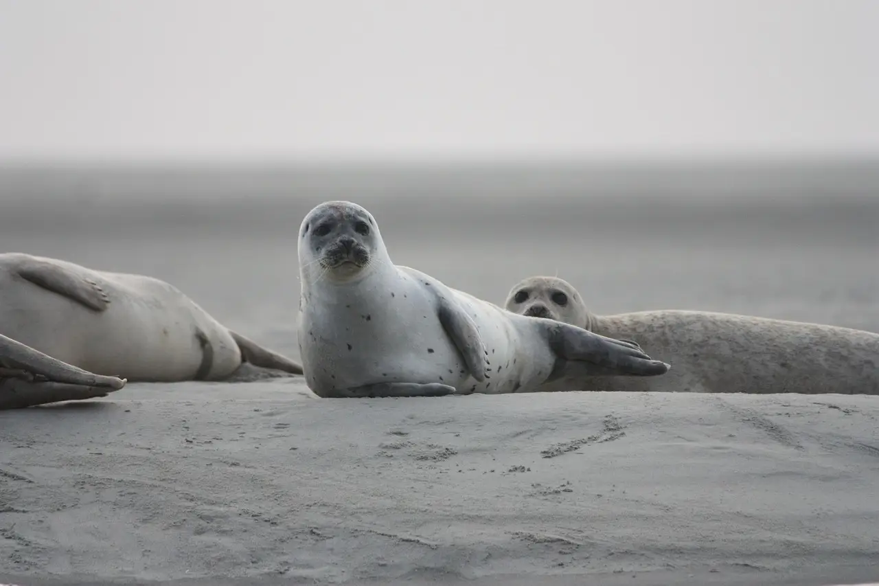
33-test neurological exam built for harbor seal pups validated in study
Veterinarians validated a 33-test neurological exam for harbor seal pups, correctly localizing lesions in all 3 suspect cases as part of research into fetal mercury exposure.
Photo: Alicia_Chant / pixabay
A 33-test neurological examination designed specifically for Pacific harbor seal pups (Phoca vitulina richardii) correctly localized lesions in all three pups with suspected neurological deficits tested, with imaging, necropsy, or both confirming each result. The protocol, published in Frontiers in Veterinary Science in 2025, gives rehabilitation clinicians a workable alternative to a land-quadruped exam that fits seals poorly.
Veterinarians developed the protocol over 2 consecutive years, examining 45 stranded pups in care at The Marine Mammal Center in Sausalito, California. After the first year, the researchers revised their draft into a 33-test exam. The final version borrows many tests from the small animal neurological examination and adds new components built around pinniped anatomy:
- a sloped ramp for evaluating proprioception and motor function
- a "handstand" for checking neck reflexes
- a fish head dangled above the pup to test balance, strength, coordination, and cranial nerve function, including the field of vision
The dangling object started as a metal nut. The team switched to a fish head after some pups tried to bite the nut. "As the harbor seals are wild animals and primarily aquatic, we had to be creative and develop new ways to assess their neurological function," Cara Field, DVM, PhD, DACZM, director of conservation medicine at The Marine Mammal Center, said in a Texas A&M news release. "Fish-head-on-a-string was a useful stimulus and, apparently, quite tasty."
How reliable is the exam?
To check consistency, the investigators repeated the exam on 17 pups. Of the 33 tests, 26 gave a dependable result in 80% or more of repeat examinations. Some pups' temperaments made certain results difficult to interpret. The authors suggested the protocol could be adapted for other phocid seals.
The study's case reports show how the exam separates neurological disease from problems that mimic it. In one pup with poor balance and coordination, MRI showed middle ear fluid consistent with otitis media plus a spinal cord lesion at C1-C2; either finding may have affected balance, and the pup improved with antimicrobial treatment and was released. Imaging revealed porencephaly in a second pup and severe hydrocephalus in a third. Three pups that seemed weak on repeat exams had primary malnutrition, with deficits the authors considered secondary to systemic illness; all three recovered enough for release.
"By developing a bespoke neurological examination protocol, we can identify pups with neurological dysfunction and pursue further diagnostic tests to identify the cause, such as intoxication, meningitis, or developmental problems," said Christine E. Thomson, BVSc, PhD, DACVIM (Neurology), DECVN, the study's first author and a veterinary neurologist at Animal Referral Hospital in Brisbane, Australia. "With a diagnosis, appropriate treatment and rehabilitation can be undertaken." The takeaway: the exam turns vague neurologic signs into anatomic targets.
What does mercury have to do with it?
The exam is one piece of a research program led by Todd O'Hara, DVM, PhD, DABVT, a veterinary toxicologist and professor at the Texas A&M University College of Veterinary Medicine and Biomedical Sciences. Mercury biomagnifies in marine food webs, building to higher concentrations at each step from prey to predator, so fish-eating marine mammals accumulate greater exposure. Contaminants also reach pups from the mother during pregnancy and through milk, making young animals a window onto very early exposures.
A 2021 study in Science of the Total Environment, from the same center, measured total mercury in blood and hair from 267 harbor seal pups admitted between 2015 and 2019, partly lanugo coat grown in utero. Mercury concentrations in both blood and hair were negatively correlated with pups' responses to touch and with movement, in both intensive care and pool stages, and higher concentrations linked to longer rehabilitation stays. The associations were modest but statistically significant, with wide individual variation and no relationship to survival.
"These findings supported that fetal exposure to mercury can put wild animals at risk for neurodevelopmental abnormalities," said Marianne Lian, DVM, PhD, that study's first author and a former doctoral student of O'Hara's. "Measuring these adverse effects can be especially difficult in a wild animal." In short: fetal mercury leaves measurable behavioral fingerprints.
What changes for rehabilitation and release?
O'Hara's group previously studied Steller sea lion pups in Alaska, but remote rookeries made thorough neurological exams hard to perform, and rehabilitated harbor seal pups offered a more workable model. "An animal may appear healthy while still having subtle neurological abnormalities that could affect its ability to survive after release," O'Hara said. "Being able to identify those effects gives rehabilitation organizations, such as the Marine Mammal Center, a more complete picture of the animal's health and whether it is ready to successfully return to the wild."
O'Hara frames the work through a One Health lens: people and marine mammals draw on many of the same marine food sources, so contaminant studies may reveal risks shared across species. Next steps include studies of California sea lions in Mexico and continued community-based work on harbor seals in Alaska, centered on mercury and other trace elements.
Source: dvm360.com
Elena Vasquez
Show full bio
Market editor covering money, funding and market shifts around Small- and large-animal clinical practice at The Vet Scope.
Related articles
Euthanasia Protocols for Exotic Pets: What the Evidence Shows
Proprioceptive deficits found in 1 of 4 dogs without neurologic signs
AAHA publishes first-ever guidelines for canine cognitive dysfunction
Diagnostic Criteria for Canine Dementia Defined Under Olby's Leadership
Wild Blacktip Sharks Hear Low-Frequency Sound From 243 Feet, Study Finds